
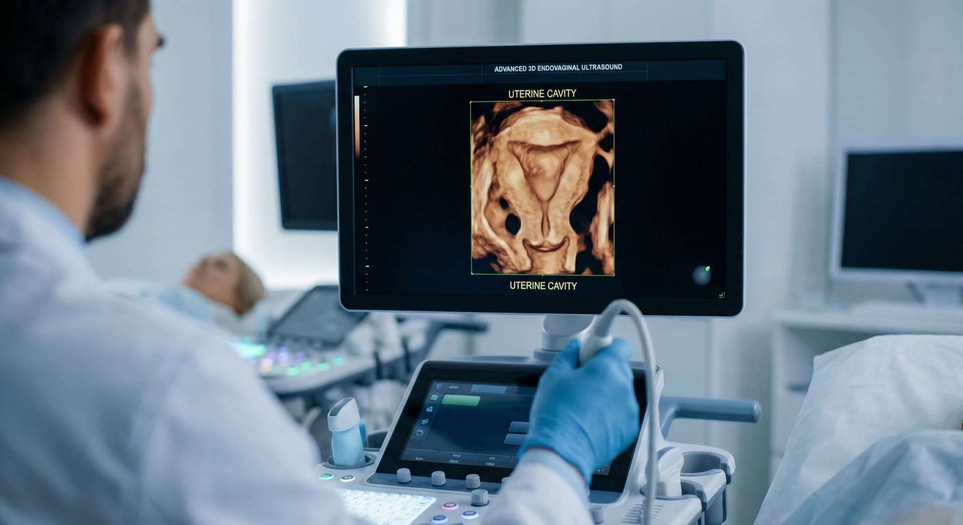


During the assessment of infertility in women, ultrasound is the preferred initial imaging method following a detailed medical history and physical examination. Ultrasound provides a basic assessment of the ovaries, the structure of the uterus, and the detection of uterine fibroids and congenital abnormalities of the uterus.
The use of three-dimensional ultrasound (3D-US), which can be employed for advanced gynaecological ultrasound assessment, is particularly crucial for the evaluation of congenital uterine anomalies, as the type and severity of these anomalies can increase pregnancy-related risks such as preterm birth and foetal positioning problems, and some of these anomalies may require surgical intervention. Three-dimensional ultrasound, which can be used for this purpose, allows for the assessment of the uterus, the uterine cavity and the simultaneous evaluation of the outer uterine borders, thereby providing guidance in clarifying the diagnosis and determining the surgical approach. Similarly, as part of an advanced gynaecological ultrasound examination, the patency of the fallopian tubes can be assessed by ultrasound following the application of gel-foam into the uterine cavity.