
PGT-M (for Single-Gene/Monogenic Disorders)

PGT-M (for Single-Gene/Monogenic Disorders)

Genetic diagnosis for the detection of single-gene disorders in the embryo.

3 min read

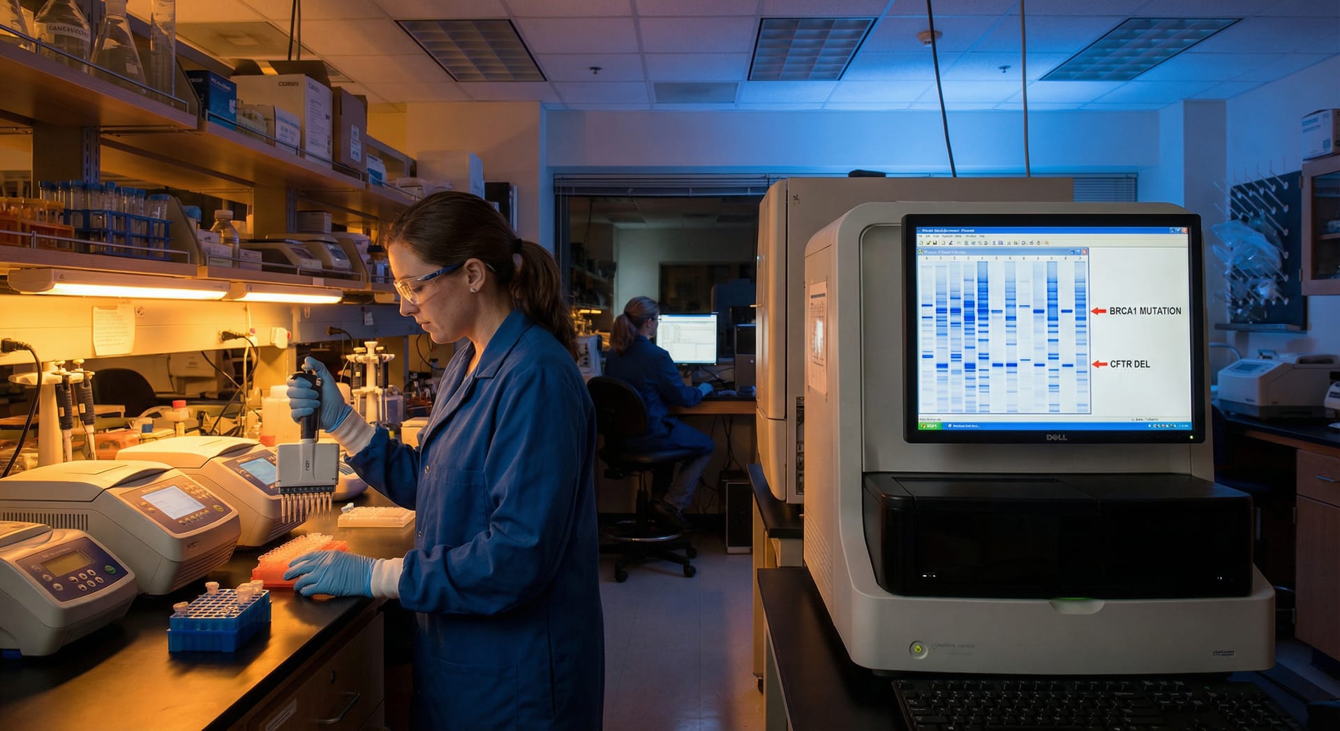
Preimplantation genetic testing for monogenic disorders (PGT-M) is a diagnostic test performed to identify healthy embryos that do not carry a genetic disorder when a hereditary condition has been identified in the family. Genetic testing of embryos is a procedure performed after the fertilisation and division process of the embryo has been completed in couples undergoing IVF treatment. In this test, a few cells, believed to represent the embryo, are taken from the embryo's outer cell mass and sent to the genetic laboratory for analysis.

PGT-M is most commonly used for conditions that present in childhood and have a severe and fatal course; in certain specific cases, it may also be used for conditions that present in adults and have a severe and serious course for which there is no treatment. In such cases, the biopsy samples taken are analysed and reported as healthy, carrier, or affected. Following the results, the transfer of a healthy embryo is prioritised; however, in cases where the embryo is reported as a carrier, appropriate genetic counselling is sought to review the options for subsequent stages.

Contact Us for More Information and Appointments

Our team of specialists will contact you as soon as possible to provide detailed information.

IVF Process

IVF treatment consists of four stages. These stages can be listed as follows: 1. Assessment of the couple, examination, diagnostic tests and determination of the treatment protocol: During the assessment of the couple, the patient's symptoms, medical history and family history are reviewed, and factors related to the cause of infertility are investigated. An ultrasound scan is performed to examine the structure of the reproductive organs, the uterus and the ovaries, and to identify any diseases or changes in neighbouring organs, investigating their relationship to infertility. If a problem is identified that hinders treatment or reduces the chances of success, steps are taken to resolve this issue first. During the ultrasound scan, an assessment of 'ovarian reserve' is carried out, which plays a significant role in selecting the treatment to be administered in the future and in determining the likelihood of treatment success. In addition, with a view to preparing for pregnancy, the presence of certain hormonal, metabolic and viral diseases that could adversely affect pregnancy, as well as the patient's immune status, is investigated. 2. Stimulation of the ovaries with medication: This constitutes the most important stage of the treatment. The treatment regimen to be administered during ovarian stimulation is determined based on the patient's age, the appearance of the ovaries on ultrasound, hormone test results, and, in patients who have previously undergone IVF, the response to treatment in the previous cycle. The physician's experience in determining this treatment directly influences the outcome. 3. Egg retrieval: Under anaesthesia, guided by transvaginal ultrasound, eggs are aspirated from the follicles containing the egg cells using a fine needle and removed from the body. 4. Embryo transfer: The best 1 or 2 embryos (fertilised eggs) are selected from those monitored in the laboratory and transferred into the uterus using a thin catheter. This procedure is similar to a routine examination and does not require anaesthesia.

The ideal time to start the egg stimulation process in IVF treatment is on the second or third day of the period. However, this is not an absolute requirement. Under certain specific conditions, ovarian stimulation medication can be started at any stage of the menstrual cycle.

Ovarian stimulation typically lasts approximately 8 to 14 days, although in some cases it may be shorter or longer. The growth of follicles that may contain eggs is monitored via ultrasound. Monitoring will require 4-5 visits to the IVF clinic during the course of treatment. In addition to ultrasound scans, hormone monitoring may also be carried out in some women. These hormones include oestrogen, progesterone and LH, and in some cases FSH. Once the follicles have reached the desired size, maturation injections (known as HCG or GnRH analogues) are administered to bring the eggs inside them to full maturity, and, barring certain special circumstances, the oocyte pick-up procedure is usually performed 34-38 hours later.

The oocyte pick-up (OPU) procedure is usually performed under local anaesthesia and takes approximately 15-30 minutes, depending on the number of follicles. If the ovarian response is limited and a small number of eggs are anticipated, the procedure may also be performed under local anaesthesia whilst the patient is awake. Following the procedure, you can return home after approximately 1-2 hours of observation and monitoring.

Once the oocyte pick-up has been completed, fertilisation with sperm takes place. The embryos are then monitored in the laboratory in special culture media for 3-5 days, or in some cases for 6-7 days. If there are any remaining high-quality embryos suitable for freezing after the transfer of those assessed in the laboratory and selected for transfer, these can be frozen and stored for transfer at a later date.

In Türkiye, the number of embryos that can be transferred is determined by regulations; for women under 35, it is limited to 1 embryo for the first 2 attempts, and for women over 35, it is limited to a maximum of 2 embryos. Under no circumstances is the transfer of more than two embryos permitted.

During the IVF treatment, ultrasound scans and blood tests are carried out during the egg stimulation phase to monitor egg development and adjust the dosage. On average, you will need to visit the hospital four or five times during the treatment process for these check-ups. As the time spent at the hospital during these check-ups is brief, they can be carried out without disrupting daily life or work routines.

Egg and Sperm Quality

Various vitamins, hormones and supplements aimed at improving egg quality have been used over the years, but none have been observed to make a clinically significant difference. Quitting smoking and losing excess weight may be beneficial. The most important factor determining egg quality is the woman's age. However, with optimal treatment, care should be taken to obtain the best and healthiest eggs from the ovaries.

Sperm quality is assessed based on three parameters: count, motility and morphological characteristics. Sperm morphology should not be considered as a separate parameter from sperm count and motility. There are certain specific structural abnormalities in sperm that can affect fertilisation and embryo development. In such cases of morphological abnormalities, sperm count and motility are also adversely affected. In such cases, specific solutions that support sperm motility and assist in the activation of the egg can be used to maintain fertilisation rates and prevent adverse effects on embryo development. It has also been demonstrated that microfluidic sperm separation methods may be beneficial. However, their superiority over conventional sperm sorting methods is debatable. Selecting sperm under high magnification (IMSI) is another method that can be used.

In vitro fertilisation, or conventional IVF, involves combining eggs retrieved from the woman with sperm retrieved from the man in a laboratory environment and transferring the embryo into the woman's uterus a certain time after fertilisation has taken place. Intracytoplasmic sperm injection (ICSI) or microinjection, on the other hand, refers to the procedure in which sperm obtained from the man is injected directly into the egg, followed by the embryo being transferred into the woman's uterus after a certain period following fertilisation. In both procedures, the primary aim is to bring the reproductive cells together in an external environment and achieve fertilisation. In the IVF method, sperm and eggs retrieved from the body are brought together in a special environment in the laboratory, and it is expected that fertilisation will occur naturally within a certain period of time. Sperm with insufficient motility and fertilisation capacity, or those that are few in number and severely malformed, cannot penetrate the egg on their own to achieve fertilisation. In such cases, fertilisation is achieved by injecting the sperm into the egg using a fine needle. This procedure is known as microinjection.

Embryo quality is assessed based on the rate of development, cell division characteristics observed during the three- to five-day monitoring period in the laboratory, and the morphological characteristics present at the blastocyst stage. A video recording of each embryo's five-day development within the incubator is examined to determine the time taken to reach specific developmental stages and the quality of these stages. This information is evaluated alongside the morphological characteristics the embryo has reached by day 5 (the structure of the inner cell mass and trophoectoderm cells, the width of the blastocyst cavity, the condition of the zona pellucida membrane surrounding the blastocyst, etc.) to determine embryo quality and select the embryo to be transferred.

Embryo Freezing (Cryopreservation) and Genetic Screening

In theory, it can be said that embryos can be preserved indefinitely after the freezing process. However, according to the IVF Regulations, once five years have elapsed following the freezing process, the preservation period for embryos can be extended by submitting a written request to the Ministry of Health.

Embryos are frozen using solutions known as specially formulated media. Nowadays, following the widespread adoption of specially prepared solutions and a rapid freezing technique known as vitrification, embryos are safely preserved in liquid nitrogen tanks at -180°C. Whereas the likelihood of embryos being adversely affected by the 'slow freezing' technique was previously 20-30 percent, this rate is now below 1 percent with the vitrification technique. However, in some cases, it may be necessary to thaw and refreeze embryos. Studies indicate that pregnancy rates may decrease after more than two cycles of freezing and thawing.

As the aim of IVF treatment is to stimulate the growth of a large number of follicles, hormone levels in the blood are higher than during a natural menstrual cycle. It is currently a matter of debate whether this may lead to the uterine lining (endometrium) maturing earlier and, consequently, whether pregnancy rates may be slightly lower. The aim is to perform a fresh transfer provided that oestrogen and progesterone hormone levels do not exceed critical thresholds. No significant difference has been found between the success rates of fresh embryo transfer and frozen embryo transfer. Scientific research indicates that pregnancy rates are similar in both cases. Therefore, fresh embryo transfer may be preferred unless there is a medical condition requiring all of the patient's embryos to be frozen.

One of the most controversial aspects of IVF treatment is the genetic screening process. In some countries, PGT-A is directly recommended and performed for patients aged 35 and over. However, as the technology still has some grey areas (mosaic embryos) and due to the possibility that embryos capable of resulting in a healthy birth may not be transferred due to false positives, embryo genetic screening is important in the following scenarios: • Older patients • Patients with a history of multiple failed attempts • Patients with chromosomal abnormalities in either the woman or the man, where there is a high likelihood of these being passed on to the embryo • Inherited single-gene disorders • Patients with unexplained recurrent pregnancy loss

In order to carry out genetic screening, it is usually necessary to remove a few cells from the embryo, which is believed to represent the embryo, once it has reached the blastocyst stage. This procedure can be performed mechanically, using an acidic chemical, or with a laser. Nowadays, this procedure can only be performed very precisely using a laser. More importantly, the embryo is allowed to reach the blastocyst stage, and a sample is taken from the trophoectoderm cells, which will form the placenta, without touching the inner cell mass, which consists of the cells that will form the embryo. Therefore, there is a possibility of the embryo being damaged during the procedure, but this risk is negligible.